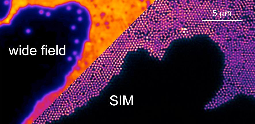
The inventors of single molecule localisation and stimulated emission depletion (STED) super-resolution microscopy were today honoured with the Nobel prize for chemistry. One of the key applications of super-resolution imaging, pioneered by the Laser Analytics Group and mentioned by the Nobel Assembly, is the study of protein aggregation reactions in the context of neurodegenerative diseases.
Super-resolution fluorescence microscopy (or nanoscopy) is a key enabler for research conducted by the Laser Analytics Group and we were thrilled to hear today that the inventors of optical nanoscopy have been honoured with the Nobel Prize.
Our group has pioneered the use of single molecule localisation microscopy, for the development of which Dr. Eric Betzig and Professor William Moerner were honoured today, in the study of protein aggregation reactions that cause neurodegeneration. We were the first to apply the technique to visualise, directly within cells that go wrong in the brains of patients suffering Alzheimer's or Parkinson's disease, how proteins called amyloids aggregate and propagate from cell to cell. The technique was first described by us in the Journal JACS in 2011 and visualises, with unprecedented resolution and within neurones, the morphology of protein aggregates of proteins causing Alzheimer's disease. In a recent paper published in the Journal JCB, and widely commented on in the news, we have used the technique to demonstrate how the protein Tau, a hallmark protein in Alzheimer's Disease, aggregates and propagates between cells and possesses a prion like infectivity.
Optical superresolution, unlike electron microscopy, is a dynamic technique, and this has allowed us to observe directly how the aggregation process of neurotoxic proteins progresses on the molecular level. In the work described in a recent paper in Nano Letters we have shown how nanoscopy resolves, molecule by molecule, how neurotoxic protein fibrils form and grow, giving molecular level insights into protein aggregation, that may one day lead to therapeutic targets against debilitating diseases such as Alzheimer's or Parkinson's.
In what has become known as nanoscopy, scientists visualise the pathways of individual molecules inside living cells.
They can see how molecules create synapses between nerve cells in the brain; they can track proteins involved in Parkinson’s, Alzheimer’s and Huntington’s diseases as they aggregate.
The Nobel Assembly discuss work pioneered by the group whilst announcing the award
Research into neurodegeneration with optical nanoscopy, pioneered by the Laser Analytics Group, was mentioned by the Nobel assembly as a key application for super-resolution microscopy during their announcement. The group has made strategic investments in the development of bespoke superresolution microscopy technologies for neurodegenerative disease research. Apart from single molecule localisation microscopy, and structured illumination microscopy, we have also now developed a STED microscope (based upon the work of prize winner Professor Hell), with which we complement our fluorescence nanoscopy toolkit in our study of neurodegeneration.
For more information about super-resolution microscopy and how this is being applied to the study of the molecular mechanism of disease please see our recent feature on this website, that explains in more detail the activities in superresolution microscopy developments in the group.
The Laser Analytics Group congratulates Dr. Betzig, Professor Hell and Professor Moerner on their outstanding achievements.
Relevant publications
Kaminski Schierle GS, van de Linde S, Erdelyi M, Esbjörner EK, Klein T, Rees E, Bertoncini CW, Dobson CM, Sauer M, and Kaminski CF, "In Situ Measurements of the Formation and Morphology of Intracellular ß-Amyloid Fibrils by Super-Resolution Fluorescence Imaging", J. Am. Chem. Soc., 133 (33), pp 12902–12905, (2011). DOI |
Michel CH, Kumar S, Pinotsi D, Tunnacliffe A, St George-Hyslop P, Mandelkow E, Mandelkow E-M, Kaminski CF, Kaminski Schierle GS, "Extracellular Monomeric Tau is Sufficient to Initiate the Spread of Tau Pathology", J. Biol. Chem. (2014), 289: 956-967. DOI | | summary
Pinotsi D, Büll AK, Galvagnion C, Dobson CM, Kaminski-Schierle GS, Kaminski CF, "Direct Observation of Heterogeneous Amyloid Fibril Growth Kinetics via Two-Color Super-Resolution Microscopy," Nano Letters (2013), 14 (1), 339–345 DOI | | summary
Fritschi S K, Langer F, Kaeser S A, Maia L F, Portelius E, Pinotsi D, Kaminski C F, Winkler D T, Maetzler W, Keyvani K, Spitzer P, Wiltfang J, Kaminski Schierle G S, Zetterberg H, Staufenbiel M, Jucker M, "Highly potent soluble amyloid-β seeds in human Alzheimer brain but not cerebrospinal fluid," Brain (2014). awu255. DOI | | summary
Esbjörner E K, Chan F, Rees EJ, Erdelyi M, Luheshi LM, Bertoncini CW, Kaminski CF, Dobson CM, Kaminski-Schierle GS, "Direct Observations of the Formation of Amyloid β Self-Assembly in Live Cells Provide Insights into Differences in the Kinetics of Aβ(1–40) and Aβ(1–42) Aggregation," Chemistry and Biology (2014) DOI || summary
Kaminski C F, Pinotsi D, Michel C H, Kaminski Schierle G S, "Nanoscale imaging of neurotoxic proteins", SPIE NanoScience+ Engineering (2013), 91690N--91690N. DOI | | summary
