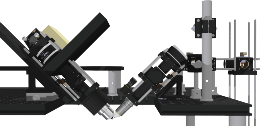
Instrumentation
We have constructed instruments that permit imaging from the molecular scale (10 nm) to whole embryos spanning 100s of microns and containing 1000s of living cells. Techniques we use include direct stochastic optical reconstruction microscopy (dSTORM) [1], structured illumination microscopy (SIM), selective plane illumination microscopy (SPIM), fluorescence lifetime imaging (FLIM) [2], [3], fluorescence anisotropy imaging microscopy (FAIM) [4], hyperspectral imaging, etc. You find more details on these instrument in the equipment overview. The technology portfolio permits us to flexibly address a large number of biological research questions. Currently we are developing variants of RESOLFT imaging with the group of Prof. Stefan Hell in Göttingen, and SPIM with Dr. Jan Felix Evers in Heidelberg, and SIM with Dr. Kevin o’Holleran at the Cambridge Advanced Imaging Centre.
Theoretical research
Our experimental research is strongly backed up by theoretical work and activities in image processing. We have made contributions to the advancement of quantitative imaging in several fields, including the global analyses of FLIM data and spectral unmixing [5], quantitative FRET assays that recover both FRET strength and stoichiometry of interactions [6], anisotropy imaging to measure rotational diffusion constants and homoFRET [7], point spread function fitting for superresolution imaging [8] [9], and single molecule diffusion trajectory imaging at superresolution. We have also performed theoretical research on frequency domain FLIM [10], which has led to the discovery of two new experimental methods to conduct FLIM, one operating at higher speed [11] and the other at better accuracy [3] than alternative FD-FLIM techniques.
FRET imaging of cyclin-cdk interactions in cells undergoing mitosis. (For details see [6].)
[1] Kaminski Schierle GS et al., "In Situ Measurements of the Formation and Morphology of Intracellular ß-Amyloid Fibrils by Super-Resolution Fluorescence Imaging" (2011)
[2] Elder AD et al., "Calibration of a wide-field frequency-domain fluorescence lifetime microscopy system using light emitting diodes as light sources" (2006)
[3] Schlacter S et al., "MhFLIM: Resolution of heterogeneous fluorescence decays in widefield lifetime microscopy" (2009)
[4] Esposito A et al., "Design and application of a confocal microscope for spectrally resolved anisotropy imaging" (2011)
[5] Schlacter S et al., "A method to unmix multiple fluorophores in microscopy images with minimal a priori information" (2009)
[6] Elder AD et al., "A quantitative protocol for dynamic measurements of protein interactions by Forster resonance energy transfer-sensitized fluorescence emission" (2009)
[7] Chan FTS et al., "HomoFRET fluorescence anisotropy imaging as a tool to study molecular self-assembly in live cells" (2011)
[8] Erdelyi M et al., "Correcting chromatic offset in multicolor super-resolution localization microscopy" (2013)
[9] Rees EJ et al., "Blind Assessment of Localisation Microscope Image Resolution" (2012)
[10] Elder A et al., "Theoretical investigation of the photon efficiency in frequency-domain fluorescence lifetime imaging microscopy" (2008)
[11] Elder AD et al., "Phi-squared fluorescence lifetime imaging" (2009)
