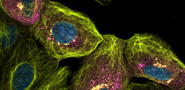
Scherer KM, Mascheroni L, Carnell GW, Wunderlich LCS, Makarchuk S, Brockhoff M, Mela I, Fernandez-Villegas A, Barysevich M, Stewart H, Suau Sans M, George CL, Lamb JR, Kaminski-Schierle GS, Heeney JL, Kaminski CF. "SARS-CoV-2 nucleocapsid protein adheres to replication organelles before viral assembly at the Golgi/ERGIC and lysosome-mediated egress" Science Advances; 8:1 (2021) DOI
Abstract
Super-resolution optical imaging reveals the accumulation of SARS-CoV-2 nucleocapsid protein at viral replication organelles. Despite being the target of extensive research efforts due to the COVID-19 (coronavirus disease 2019) pandemic, relatively little is known about the dynamics of severe acute respiratory syndrome coronavirus 2 (SARS-CoV-2) replication within cells. We investigate and characterize the tightly orchestrated virus assembly by visualizing the spatiotemporal dynamics of the four structural SARS-CoV-2 proteins at high resolution. The nucleoprotein is expressed first and accumulates around folded endoplasmic reticulum (ER) membranes in convoluted layers that contain viral RNA replication foci. We find that, of the three transmembrane proteins, the membrane protein appears at the Golgi apparatus/ER-to-Golgi intermediate compartment before the spike and envelope proteins. Relocation of a lysosome marker toward the assembly compartment and its detection in transport vesicles of viral proteins confirm an important role of lysosomes in SARS-CoV-2 egress. These data provide insights into the spatiotemporal regulation of SARS-CoV-2 assembly and refine the current understanding of SARS-CoV-2 replication.
