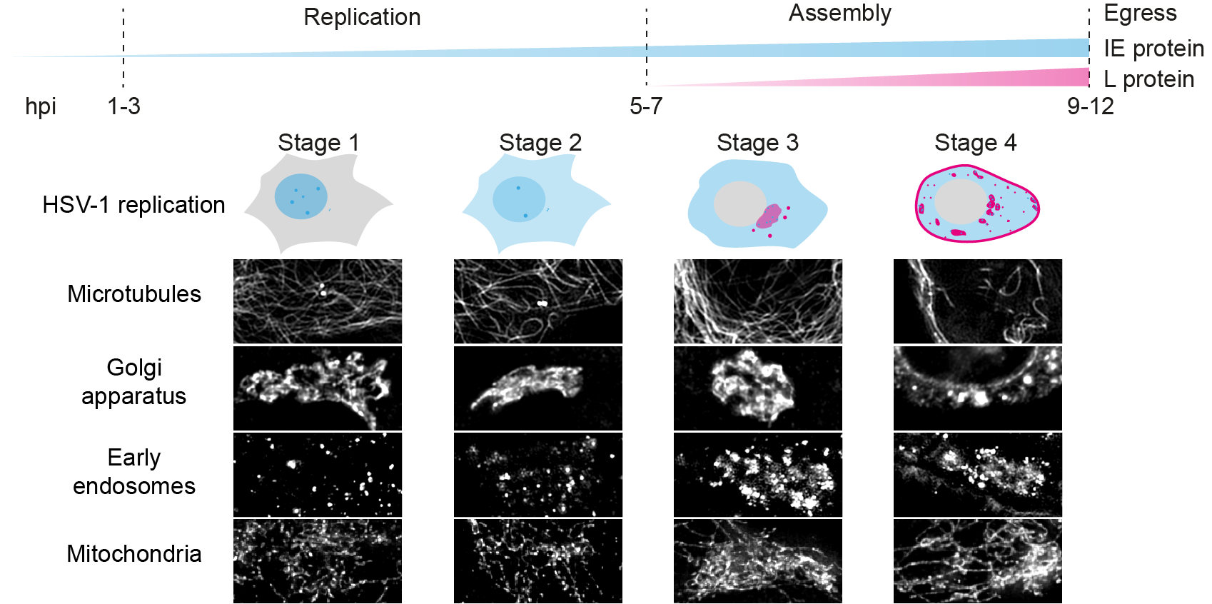
Figure: Fluorescent timestamping for the analysis of host cell remodelling during HSV-1 replication
Summary
Herpesviruses are large and complex DNA viruses that are composed of an icosahedral capsid, a proteinaceous layer termed the tegument, and a glycoprotein rich lipid envelope. One important area of host-pathogen interaction that is still poorly understood is the extensive change to intracellular organelles and cellular morphology that occur within the infected cell during active virus replication. In order to characterise the spatiotemporal dynamics of host cell remodelling caused by herpesvirus infection, we use novel multiparametric fluorescence microscopy methods compatible with live-cell imaging. In addition, we apply expansion microscopy to map 3D rearrangement in great detail. The remodelling of the host cell is correlated to the stage of virus replication which can highly vary between individual cells. Therefore, we have constructed a recombinant reporter virus that expresses eYFP-tagged ICP0, a multifunctional immediate early tegument protein, as well as mCherry-tagged glycoprotein C (gC), a late protein that is a major component of the viral envelope. The sequential expression of these two viral proteins provides us with an intrinsic time stamp for the stage of virus infection in each cell. With this fluorescent reporter virus, we are able to describe the remodeling of- the three-dimensional architecture of microtubules and the actin network,- compartments of the secretory and endocytic pathways which are intimately linked to viral envelope protein synthesis, maturation and transport,- and key antiviral and inflammatory signalling platforms (mitochondria and peroxisomes).
Scherer KM, Soh T, Manton JD, Mascheroni L, Crump CM, Kaminski CF. Spatiotemporal dynamics of host cell modification caused by herpesvirus infection. Access microbiology. (2019) DOI

