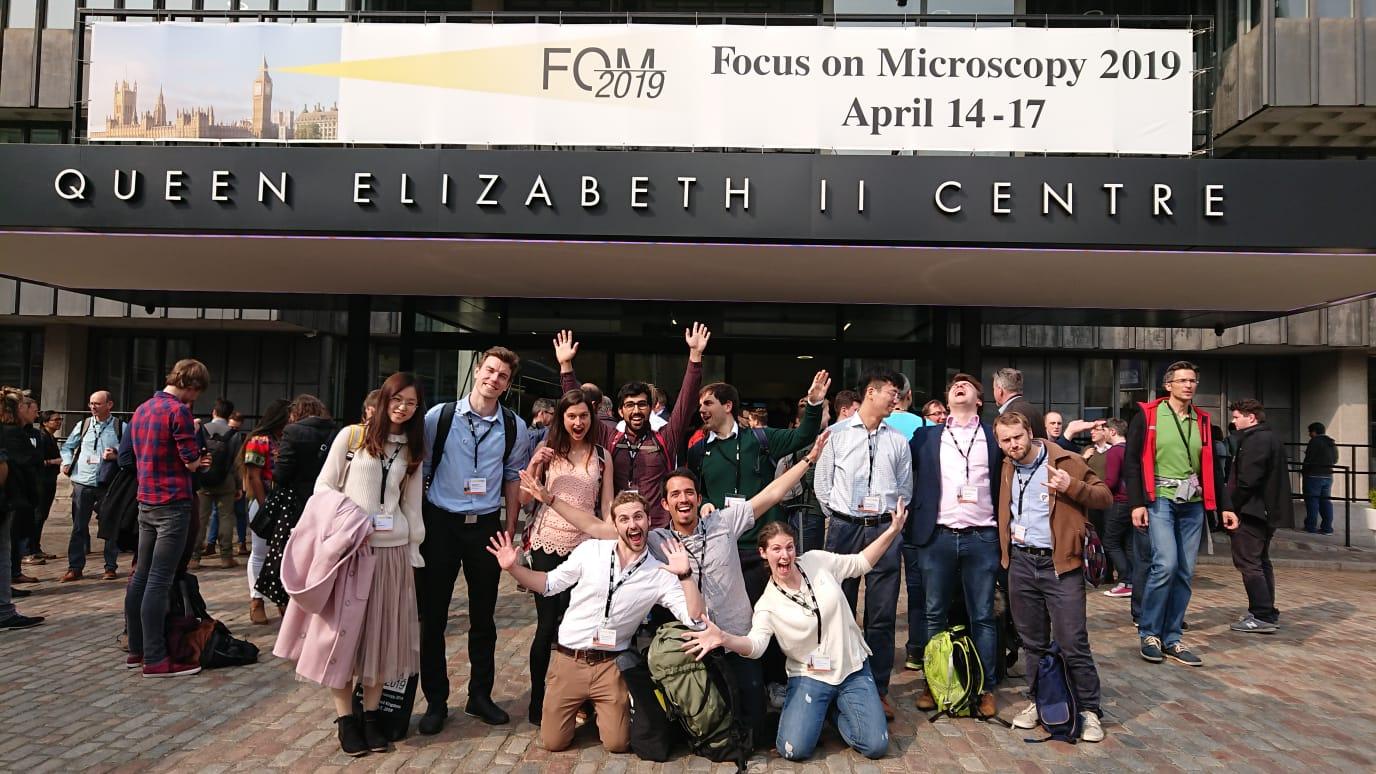
Members of the Laser Analytics Group and the Molecular Neuroscience Group attended the Focus on Microscopy Conference in London.
This year's Focus on Microscopy conference featured an array of talks and posters from current members of the Laser Analytics Group and the Molecular Neuroscience Group, listed below:
- Prof. Clemens Kaminski: Optical Superresolution Microscopy of Molecular Mechanisms of Disease (invited)
- Dr. Katharina Scherer: Spatiotemporal Dynamics of Host Cell Modification during Herpes virus Replication
- Dr. Ioanna Mela: Correlative Afm/STED and Afm/FLIM Imaging for the investigation of Mechanical and Functional Material properties
- Charles Christensen: Image Restoration Using Deep Convolutional Neural Networks Applied to High-Speed Optical Microscopy of Living Samples
- Lisa Hecker: A Novel, Cost-Efficient Concept for Interferometric Multi-Colour Structured Illumination Microscopy
- Wei Li: Fluorescent Carbon Dots for Super-Resolution Imaging
Some snazzy-looking posters were presented by
- Chetan Poudel: Fast Time-Gated FLIM Enables High-Throughput Functional Imaging of Protein Aggregation in Moving C. Elegans
- Oliver Vanderpoorten: 1-Dimensional Diffusion Processes in Two-Photon Fabricated Pdms Nanochannel Devices
- Pedro Vallejo Ramirez: A Superresolution Method to Quantify the Association of Alpha Synuclein with Synaptic Organelles in the Study of Neurodegeneration
- Lucia Wunderlich: Optical Super-Resolution Microscopy of the Kidney: a Practical Guide
- Dr. Meng Lu: Advanced Imaging of Amyloid-Beta Protein Aggregation
- Luca Mascheroni: From Fish to Viruses: Refining Expansion Microscopy for the Imaging of Diverse Biological Samples
- Miranda Robbins: Imaging Neuronal Synapses to Understand Memory Impairment in Neurodegeneration
In addition, some former LAG members presented contributing talks as well:
- Dr. Florian Strohl: Label-Free Nanoscopy Enabled by Photonic Chip Fourier Ptychography
- Dr. Romain Laine: Divide and Conquer: Combining Structured Illumination Microscopy with Machine Learning for Large Scale Structural Analysis of Viruses
- James Manton: Lorentzian Light Sheets: Longer, Thinner, More Efficient Illumination Profiles for Macroscopic Samples
- Dr. Nathan Curry: High Content 3D and 4D Oblique Plane Microscopy in 96- and 384-Well Plate Formats
- Dr. Chris Rowlands: High Speed Deep Tissue Multiphoton Brain Imaging
- Dr. Marcus Fantham: The Fpbioimage Plugin for Imagej
